Over the course of my career, some of the most satisfying—and often surprising—results I have seen in periodontal practice have come from balancing what I call the three pillars of periodontal healing: root cleanliness, control of the infectious component, and occlusion.
All three matter. But for this discussion, I want to begin with the infectious component because it raises an interesting question: If we believe we are treating an infection, shouldn’t we try to identify what we are treating?
For many years, periodontal antibiotics were largely prescribed empirically. Clinical judgment remains important, but today we have another source of information. In our practice, we use OralDNA® salivary testing in selected periodontal patients to help identify the bacterial component of the disease. Does every positive bacterial identification mean antibiotics? No. It means that we have a measure that we can compare pre-treatment to post-treatment.
Testing does not mean that every positive result requires an antibiotic. Far from it. When I believe an antibiotic may be indicated, I would rather have additional information about the organisms involved than prescribe blindly. The bacterial profile can complement the clinical examination and help inform antibiotic selection.
There is also a clinical presentation that particularly gets my attention. What we once called aggressive periodontitis often presents today within the Grade C category, and clinically it can look and feel like a different disease. During periodontal probing, the probe may seem to drop into the pocket with almost no resistance—little or no detectable calculus and a lack of the firm tissue resistance we normally encounter. We don’t see this frequently—probably in fewer than 10% of our cases—but when we do, it changes our approach.
In those patients, we test first. When indicated, we select the antibiotic based in part on the bacterial findings and then reevaluate the patient approximately one week after completion of the antibiotic, before beginning definitive periodontal treatment. If we see significant clinical improvement—and often we do—we then proceed with treatment.
That sequence gives us information. We are not simply assuming that infection is important; we have identified the organisms, treated the infectious component when appropriate, and observed the patient’s response.
But bacterial identification is only one pillar.
Another is root cleanliness. Periodontal endoscopy has been an important part of our practice for more than 25 years because it allows our hygienists to visualize the subgingival root surface and identify deposits that conventional instrumentation may leave behind. More recently, we have also incorporated EDTA to address residual microislands of calculus described by Harrel and Cobb.
The third pillar is occlusion. Fremitus matters. Occlusal forces matter. A severely compromised tooth with reduced periodontal support may also be subjected to forces it is poorly equipped to tolerate. We routinely evaluate those forces and adjust them when appropriate.
And then we wait.
That may be one of the most important parts of the entire process. Once the root has been cleaned, the infectious component addressed, and excessive forces reduced, we give the tissues time to respond before making an irreversible decision.
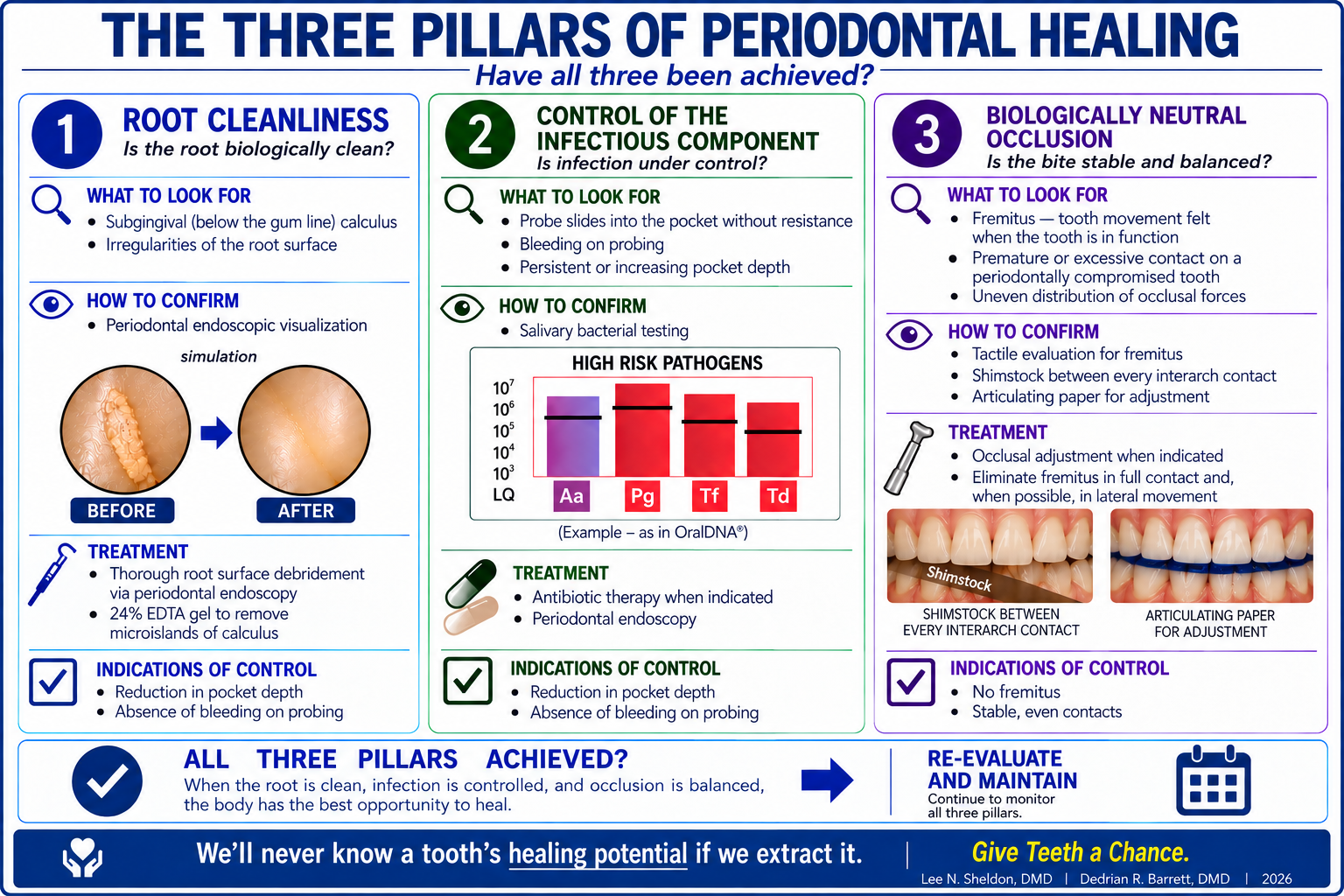
Not every severely compromised tooth can—or should—be saved. But after decades of treating periodontal disease, I have become increasingly reluctant to call a tooth hopeless until we have addressed all three pillars and given healing an opportunity to occur.
Salivary testing hasn’t replaced clinical judgment in our practice. It has given us another piece of information with which to exercise it.
And when the alternative may be extraction, more information can matter.
Once a tooth is extracted, we will never know how well it might have healed.
References
- Stambaugh RV, Dragoo M, Smith DM, Carasali L. The limits of subgingival scaling. Int J Periodontics Restorative Dent. 1981;1(5):30–41.
- Harrel SK, Wilson TG Jr, Tunnell JC, Stenberg WV. Laser identification of residual microislands of calculus and their removal with chelation. J Periodontol. 2020;91:1562–1568.
- Harrel SK, Cobb CM, Nunn ME, Zhao D. Laser and scanning electron microscopy evaluation of residual microislands of calculus. J Periodontol. 2025;96:268–278.
- Harrel SK, Cobb CM, Sheldon LN, Rethman MP, Sottosanti JS. Calculus as a risk factor for periodontal disease: a narrative review on treatment indications when the response to scaling and root planing is inadequate. Clin Exp Dent Res. 2022;8(6):1385–1394.


Dr. Sheldon’s work emphasizes the roles of calculus, biofilm, inflammation, occlusion, and patient-specific risk factors—areas that are increasingly illuminated by salivary diagnostics and precision tools such as those developed by OralDNA. He has long advocated that better diagnosis, not more invasive treatment, is often the key to long-term success.
A national lecturer and educator, Dr. Sheldon challenges extraction-first and procedure-driven models of care, encouraging dentists and hygienists to rethink how periodontal disease is identified, managed, and monitored. Now semi-retired from clinical practice, he continues to teach and write, focusing on changing how dentistry thinks about healing, prevention, and patient-centered outcomes.
- The Three Pillars of Periodontal Healing: Why Identifying the Infection Matters - October 23, 2026
- Periodontal Endoscopy: Seeing What Stops Periodontal Disease - July 10, 2026
- When a “Hopeless” Tooth Isn’t Hopeless - January 23, 2026